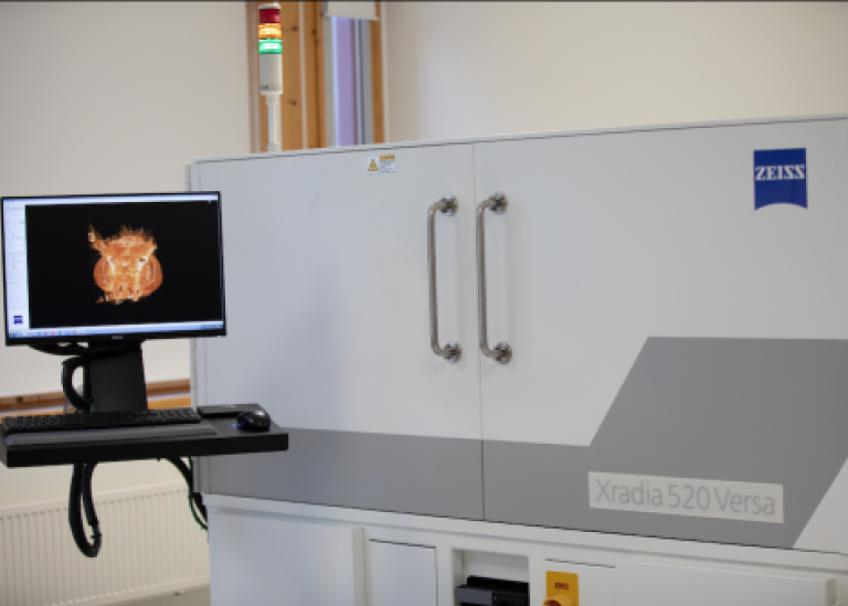
X-ray microscope: Xradia Versa 520
SUBIC has a laboratory X-ray imaging system: Zeiss Xradia Versa 520.
Zeiss Xradia Versa 520 is the most advanced model in the Xradia Versa family. With user-friendly control software, automatic loading robot, and advanced microscopic system, our machine offers the best experience for researchers. It has been used to make non-destructive, high resolution 3D Imaging of everything from insect eyes to animal bones and fossils.
Starting, running and finishing a project with X-ray
Applications and techniques
There are a variety of fields that can benefit from the X-ray technique. At SUBIC, scientists from many different research areas use this equipment. Here are a few examples:
- Life Sciences
- Medicine and biomedical research
- Materials and Engineering
- Environmental Sciences
- Physics
- Paleontology and Archeology
- Industrial applications
- Food science
Techniques
The Zeiss Xradia Versa 520 can operate with different methodologies, for example:
- Radiography: 2D imaging
- Tomography: 3D imaging
- Dual energy scan: Imaging at two different X-ray energies can potentially provide quantitative information about the composition and density of the object with multiple materials.
- Phase-contrast imaging: Using phase shift of the X-rays to enhance the image contrast for low-absorbing materials.
Instrument
X-ray source
- Microfocus tungsten X-ray source
- Energy range: 10.0 - 160.0 keV
Detector
- Scintillator-based camera with 4 different objectives: 0.4x, 4x, 20x and 40x
- CCD: 2048 x 2048 pixels, 13.5 µm pixel size
- Field of view (FOV): adjustable between approximately 0.5 mm to 50 mm. Stitching is possible to extend the FOV further.
- Effective pixel sizes adjustable between approximately 0.1 µm and 34 µm (if no binning). It depends on the objective (optical magnification) and the distances between the source, sample and detector (geometrical magnification).
- Spatial resolution: approximately 0.7 µm in 2D, about 1 µm in 3D.
Examples
Contact
For more information regarding X-ray imaging, technical equipment, or other X-ray related requests, please contact Tunhe Zhou: tunhe.zhou@su.se
Last updated: December 21, 2023
Source: Department of Linguistics/SUBIC