About the facility
IVMSU is a national infrastructure for two-photon microscopy that supports intravital and non-linear light microscopy with open access to national and international users.
On this page you can learn more about our facility. Do not hesitate to reach out to us if you have any questions.
Accessibility
Web-based application via the NMI portal http://www.nmisweden.se.
Access based on
- Feasibility
- Capacity
- Recommended general guidelines from the national steering committee
Requirement for ethical permit from local ethical committee for in vivo-studies.
For BSL2 studies: Risk assessment for biological agent and toxins.
IVMSU units
- Imaging room
- Surgery lab
- Facilities for maintaining in-vivo-models
Support and service
- Microscopy- assistance and training
- Project discussion and study design
- Surgery- assistance and training
- Support for the preparation of ethical permits
- Assistance with transfer of applications/permits to the Stockholm University facility permit
- Usage of cell lab equipment (incubator and laminar flow cabinet)
- Animal handling in quarantine facility and animal housing room
- Documentation, logging and overview of in vivo-models in database system
- Possibilities for image analysis at the nearby IFSU facilities (LAS-X and Imaris)
- Access to SU-based data storage
User fees
We support feasibility tests free of charge.
For more information about fees, please contact IVMSU@su.se.
Projects
Ongoing projects - Examples
Multicolor cell labeling for solving the clonal structure of murine embryo neural crest
Department of Medical Bioschemistry and Biophysics, Karolinska Institutet
In this project, we will experimentally target clonal structures of the cranial neural crest cells and will study their reparative plasticity as well as underlying signaling mechanisms that can be considered as new therapeutic targets. At the single cell and clonal levels, we will systematically explore the spectrum of signals integrating the growing oral and craniofacial complex.
The effect of temperature on the bumblebee brain
Department of Zoology, Stockholm University
The aim of the project is to understand if and how the information carried by neurons in the bumblebee brain is affected by temperature. The findings will have consequences for understanding how increasing environmental temperatures, such as those that are occurring as a result of climate change, will affect the function of insect nervous systems and behavior.
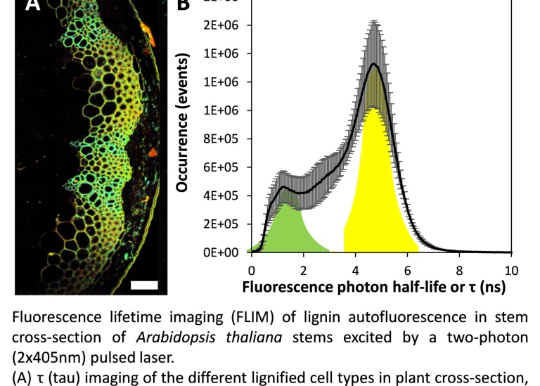
Evaluation of autofluorescence life-time imaging to observe differences in biological samples
Department of Ecology, Environment and Plant Sciences, Stockholm University
Imaging Candida albicans infection in the mouse kidney
Department of Molecular Biosciences, Stockholm University
Visualization of Toxoplasma dissemination and immune cell migration in mice
Department of Molecular Biosciences, Stockholm University

Ex vivo characterisation of the regenerating salamander heart - Department of Cell and Molecular Biology,
Karolinska Institutet
*All in vivo projects are performed in compliance with ethics permits approved by the local ethics committee.
Finished projects - Examples
- Multiphoton imaging of bacterial biofilm structures. Karolinska Institutet
- Flipper-TR plasma membrane tension. CMM, Karolinska Institutet
- FRET-FISH: combining DNA FISH with FLIM-FRET for quantification of chromosome X inactivation in mouse. Department of Physiology and Pharmacology, Karolinska Institutet
- Live monitoring of cellular dynamics during skeletal development and maintenance. Department of Physiology and Pharmacology, Karolinska Institutet.
- Second harmonic generation imaging of collagen gels. Department of Micro and Nanosystems, KTH Royal Institute of Technology, Sample: hydrogel based on type I collagen
- Determining the effect of Piezo1 activation on membrane tension in cancer cells. Department of Medical Cell Biology, Uppsala University. Samples: MDA-MB-231 breast cancer cells
- Real-time visualisation of human stem cells during neuronal differentiation. Department of Organismal Biology, Uppsala University. Samples: Human embryonic stem cells.
- Multicolor cell labeling for solving the clonal structure of murine embryo neural crest. Department of Medical Bioschemistry and Biophysics, Karolinska Institutet
- The effect of temperature on the bumblebee brain. Department of Zoology, Stockholm University
*All in vivo projects are performed in compliance with ethics permits approved by the local ethics committee.
Publications
Functional Complexity on a Cellular Scale: Why In Situ Analyses Are Indispensable for Our Understanding of Lignified Tissues, Blaschek L, Serk H, Pesquet E. Functional Complexity on a Cellular Scale: Why In Situ Analyses Are Indispensable for Our Understanding of Lignified Tissues. J Agric Food Chem. 2024 Jun 4. doi: 10.1021/acs.jafc.4c01999. Epub ahead of print. PMID: 38832924.
Bulk and In Situ Quantification of Coniferaldehyde Residues in Lignin, Pesquet E, Blaschek L, Takahashi J, Yamamoto M, Champagne A, Nuoendagula, Subbotina E, Dimotakis C, Bacisk Z, Kajita S. Bulk and In Situ Quantification of Coniferaldehyde Residues in Lignin. Methods Mol Biol. 2024;2722:201-226. doi: 10.1007/978-1-0716-3477-6_14. PMID: 37897609.
The maturation of native uropathogenic Escherichia coli biofilms seen through a non-interventional lens, Zhang T, Ray S, Melican K, Richter-Dahlfors A. The maturation of native uropathogenic Escherichia coli biofilms seen through a non-interventional lens. Biofilm. 2024 Jul 6;8:100212. doi: 10.1016/j.bioflm.2024.100212. PMID: 39114648; PMCID: PMC11305213.
Blue-shift photoconversion of near-infrared fluorescent proteins for labeling and tracking in living cells and organisms, Pennacchietti F, Alvelid J, Morales RA, Damenti M, Ollech D, Oliinyk OS, Shcherbakova DM, Villablanca EJ, Verkhusha VV, Testa I. Blue-shift photoconversion of near-infrared fluorescent proteins for labeling and tracking in living cells and organisms. Nat Commun. 2023 Dec 19;14(1):8402. doi: 10.1038/s41467-023-44054-9. PMID: 38114484; PMCID: PMC10730883.
Proline catabolism is a key factor facilitating Candida albicans pathogenicity, Silao FGS, Jiang T, Bereczky-Veress B, Kühbacher A, Ryman K, Uwamohoro N, et al. (2023) Proline catabolism is a key factor facilitating Candida albicans pathogenicity. PLoS Pathog 19(11): e1011677. https://doi.org/10.1371/journal.ppat.1011677
Blood-brain barrier-restricted translocation of Toxoplasma gondii from cortical capillaries. Gabriela C. Olivera, Emily C. Ross, Christiane Peuckert, Antonio Barragan. Elife. 2021;10:e69182.doi: 10.7554/eLife.69182.
Three Dimensional Microvascularized Tissue Models by Laser-Based Cavitation Molding of Collagen. Alessandro Enrico, Dimitrios Voulgaris, Rebecca Östmans, Naveen Sundaravadivel, Lucille Moutaux, Aurélie Cordier, Frank Niklaus, Anna Herland, and Göran Stemme. Adv Mater. 2022;e2109823.doi: 10.1002/adma.202109823
FRET-FISH probes chromatin compaction at individual genomic loci in single cells. Mota A, Berezicki S, Wernersson E, Harbers L, Li-Wang X, Gradin K, Peuckert C, Crosetto N, Bienko M. Nat Commun. 2022 Nov 5;13(1):6680. doi: 10.1038/s41467-022-34183-y.
Last updated: October 8, 2025
Source: MBW