Research project Unraveling the local structure of Cellulose Nanocrystals using Scanning Electron Diffraction
Cellulose is an essential constituent in the architecture of plants, where it contributes with extraordinary properties in terms of strength and stiffness. Using scanning electron diffraction we are revealing details of its atomic structure.
The aim of this project is to open up for the development of new materials based on cellulose through the generation of new understanding about its local crystalline structure at the nanoscale. The impressive mechanical properties of cellulose are believed to originate from its crystal structure and the complex pattern of covalent and hydrogen bonding. In this project, we are using an emerging method in the transmission electron microscope called scanning electron diffraction (SED). These data will provide insights to the packing of the individual polysaccharide chains in the cellulose nanofibers which will help to understand their properties.
Project description
Plants form an impressive variety of structural architectures. In order to make this possible, nature has developed a complex multidimensional material, which exhibits extraordinary mechanical properties. Wood is an abundant source of cellulose in Sweden and worldwide. By acid hydrolysis or TEMPO oxidation combined with mechanical processing, the principal cellulose fibers can be isolated from its natural sources and be a biomaterials resource to construct hybrid materials.
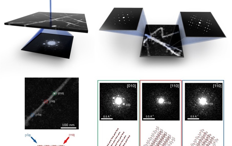
In this project, we are building an increased knowledge about the structures at the atomic scale of these biopolymeric materials. In order to achieve this we are using emerging methods in the scanning transmission electron microscope (STEM), e.g. scanning electron diffraction (4D STEM. By scanning a nanometer-sized electron probe across the sample and acquiring a 2D electron diffraction pattern for each raster point, a 4-dimensional data set can be constructed. The data is acquired using very fast detectors, which can acquire several thousand patterns per second. Using this method, a map revealing the ordering of atoms in the sample with nanometer resolution is created.
With a deeper understanding of the local crystal structure of cellulose, we will contribute to the understanding of the relationship between structure and properties of the cellulose nanofibers. More profound knowledge about this will lead to better ways to process and extract the CNCs from their natural sources as well as tailoring their properties, opening up for the development of materials with new and improved properties.
Project members
Project managers
Tom Willhammar
Associate Professor

Members
Mathias Nero
PhD Student