Håkan FischerProfessor i Humanbiologisk psykologi
Om mig
Sedan 2011 har jag varit professor i humanbiologisk psykologi och chef för avdelningen för Psykobiologi och epidemiologi vid Psykologiska institutionen, Stockholms universitet. Jag var prefekt vid Psykologiska institutionen mellan 2015 och 2021. Dessutom är jag docent vid Karolinska Institutet (KI), knuten till Aging Research Center vid KI och Stockholms universitet Brain Imaging Centre (SUBIC). Jag är också fakultetsmedlem vid Digital Futures, ett tvärvetenskapligt forskningscenter vid Kungliga Tekniska Högskolan i Stockholm.
Jag har en doktorsexamen i psykologi från Uppsala Universitet sedan 1998. Från 1999 till 2001 var jag postdoktoral forskare vid Harvard Medical School i Boston, USA. Därefter arbetade jag som forskare vid Aging Research Center, KI, innan jag började vid Psykologiska institutionen vid Stockholms universitet 2011. Jag tillbringade läsåret 2021–2022 på sabbatsår vid University of Florida, Department of Psychology.
Nuvarande positioner och roller
- Chef för avdelningen för Psykobiologi och epidemiologi (tidigare Biologisk psykologi), Psykologiska institutionen, Stockholms universitet (2011–nuvarande)
- Chef för enheten för Kognitiv neurovetenskap, Psykologiska institutionen, Stockholms universitet (2024–nuvarande)
- Ledamot i Vetenskapsrådets ämnesråd för humaniora och samhällsvetenskap (2023–nuvarande)
- Stockholms universitets representant i styrgruppen för EBRAINS Sweden (2022–nuvarande)
- Stockholms universitets representant i styrgruppen för samarbetet mellan Stockholm Trio och University of Tokyo, Japan (2022–nuvarande)
- Ledamot i styrgruppen för samarbetet mellan Stockholms universitet och RISE (Research Institutes of Sweden) (2019–nuvarande)
- Representant i forskningsutvärderingsgruppen för humaniora och samhällsvetenskap vid Stockholms universitet (2021–nuvarande)
- Huvudforskare i spin-off företaget Empatik AI/Aiyo AB (2022–nuvarande)
Undervisning
Jag är kursledare för grundkursen Kognitiv neurovetenskap och masterkursen Emotionspsykologi och affektiv neurovetenskap.
Jag undervisar regelbundet på både grund- och avancerad nivå, främst inom biologisk psykologi, kognitiv neurovetenskap och emotionspsykologi, med fokus på samspelet mellan hjärna och beteende.
För närvarande är jag huvudhandledare för en doktorand och biträdande handledare för fyra doktorander.
Forskning
Mitt huvudsakliga forskningsområde är att undersöka intra- och interindividuella skillnader i affektiv, kognitiv, social och perceptuell bearbetning, samt relationen mellan dessa beteenden och hjärnfunktion, med ett specifikt fokus på åldersskillnader hos vuxna. Målet är nu att använda single-subject small-N designs och variabilitet för att studera mekanismerna bakom dessa processer.
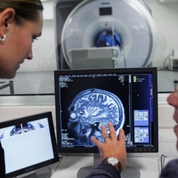
För att studera funktionell hjärnaktivering använder jag funktionell magnetresonanstomografi (fMRI), positronemissionstomografi (PET) och funktionell nära-infraröd spektroskopi (fNIRS). För att studera hjärnstruktur använder jag T1-viktad avbildning, diffusionstensoravbildning (DTI) och perfusionsavbildning. Jag samarbetar både nationellt och internationellt med andra forskare och är involverad i pågående projekt i Sverige, Tyskland och USA.
Jag deltar i nio pågående finansierade forskningsprojekt, två som huvudansvarig (totalt 6,9 miljoner SEK) och sju som medsökande (totalt 24,8 miljoner SEK).
Nuvarande forskningslinjer
- Intra- och interindividuella skillnader i perception och igenkänning av socio-emotionell information: Tillsammans med forskare vid Stockholms universitet (SU), Uppsala Universitet (UU), Karolinska Institutet (KI) och University of Florida (UF), studerar vi förmågan att upptäcka socio-emotionell information.
- Effekten av oxytocin på socio-emotionell informationsbearbetning: Undersökning av den neurobiologiska grunden för oxytocins effekter på socio-emotionell bearbetning hos yngre och äldre vuxna, i samarbete med forskare vid SU, UU, KI, UF och Göteborgs universitet.
- Utveckling av artificiell intelligens (AI) för interpersonell kommunikation: Samarbetar med forskare vid SU, UU, UF och Institutionen för data- och systemvetenskap, SU, för att stödja läsning, tolkning och förutsägelse av interpersonella kommunikationsmönster.
Handledning och finansiering
För närvarande handleder jag sex doktorander. Sedan 2002 har jag konsekvent erhållit finansiering från olika prestigefyllda källor, inklusive Vetenskapsrådet, Wallenbergstiftelsen (MMW och MAW), Stiftelsen för internationalisering av högre utbildning och forskning (STINT), Riksbankens Jubileumsfond och Konung Gustav V och Drottning Victorias stiftelse.
Publikationer och akademiska bidrag
Mitt vetenskapliga arbete har resulterat i 136 forskningsartiklar publicerade i eller inskickade till peer-reviewed tidskrifter. Mina forskningspublikationer har citerats mer än 12 000 gånger i internationella vetenskapliga tidskrifter och har ett h-index (Google) på 52 och ett i10-index på 116. Jag har även suttit i flera halvtids- och doktorsexamenskommittéer, bedömt forskning och tjänstetillsättningar vid olika akademiska institutioner samt varit granskare för flera internationella peer-reviewed tidskrifter.
Manuskript under granskning och revidering
- Fischer, H., Collier, E.S., Harris, K.L., Manzouri, A., Skedung, L., Rutland, M.W. Active Touch in Tactile Perception: Brain Activity and Behavioral Responses to Surface Differences. SSRN Paper (preprint), accepterad i tidskriften Experimental Brain Research.
Inslag i media (urval på svenska)
- Allt du velat veta: Om kärlekens kemi med Håkan Fischer – vad händer i hjärnan när vi blir förälskade?
- I hjärnan på Louise Epstein: Det händer i hjärnan när du blir kär
- Forskarliv P1: Forskarliv
- Nyhet om forskning P1: Nyhet om forskning
- Stockholms universitets webb: AI som ska kunna läsa av dina känslor
Inslag I media (urval på engelska)
Forskningsprojekt


 av Nora Choque Olsson.")

Publikationer
I urval från Stockholms universitets publikationsdatabas
-
Associations between Genetic Variations in Oxytocin Pathway Genes and Hippocampal Volume: Insights from the UK Biobank
2025. Shanshan Xiao (et al.). Cortex
ArtikelLäs mer om Associations between Genetic Variations in Oxytocin Pathway Genes and Hippocampal VolumeThe role of oxytocin-related genes in social-cognitive function has been previously established, but structural brain mechanisms underlying this link remain poorly understood. Utilizing a substantial dataset from the UK Biobank (N ≈ 30,000), this research determined associations between variations in ten single nucleotide polymorphisms (SNPs) within three oxytocin pathway genes (i.e., the oxytocin/neurophysin I prepropetide gene, the cluster of differentiation 38 glycoprotein gene, the oxytocin receptor gene) and whole-brain gray matter volume. Carriers of the AA or AG genotypes of the oxytocin receptor gene rs237851 SNP exhibited significantly larger hippocampal volume than carriers of the GG genotype. These results support the link between variations in the oxytocin receptor gene and hippocampal structure, with possible impact on social-cognitive function such as social recognition memory.
-
Altered Empathy Processing in Frontotemporal Dementia
2024. Olof Lindberg (et al.). JAMA Network Open 7 (12)
ArtikelLäs mer om Altered Empathy Processing in Frontotemporal DementiaIntroduction: Loss of empathy is a core symptom of behavioral variant frontotemporal dementia (bvFTD).1 In particular, the affective aspect of empathy appears to be independent of decrease in the other socioemotional abilities and general cognition in bvFTD.2We used an established functional magnetic resonance imaging (MRI) paradigm3 to assess bvFTD-related alterations in brain responses during empathy for pain (EFP) in a case-control study.
-
Effects of four-week intranasal oxytocin administration on large-scale brain networks in older adults
2024. Peiwei Liu (et al.). Neuropharmacology 260
ArtikelLäs mer om Effects of four-week intranasal oxytocin administration on large-scale brain networks in older adultsOxytocin (OT) is a crucial modulator of social cognition and behavior. Previous work primarily examined effects of acute intranasal oxytocin administration (IN-OT) in younger males on isolated brain regions. Not well understood are (i) chronic IN-OT effects, (ii) in older adults, (iii) on large-scale brain networks, representative of OT's wider-ranging brain mechanisms. To address these research gaps, 60 generally healthy older adults (mean age = 70.12 years, range = 55–83) were randomly assigned to self-administer either IN-OT or placebo twice daily via nasal spray over four weeks. Chronic IN-OT reduced resting-state functional connectivity (rs-FC) of both the right insula and the left middle cingulate cortex with the salience network but enhanced rs-FC of the left medial prefrontal cortex with the default mode network as well as the left thalamus with the basal ganglia–thalamus network. No significant chronic IN-OT effects were observed for between-network rs-FC. However, chronic IN-OT increased selective rs-FC of the basal ganglia–thalamus network with the salience network and the default mode network, indicative of more specialized, efficient communication between these networks. Directly comparing chronic vs. acute IN-OT, reduced rs-FC of the right insula with the salience network and between the default mode network and the basal ganglia–thalamus network, and greater selective rs-FC of the salience network with the default mode network and the basal ganglia–thalamus network, were more pronounced after chronic than acute IN-OT. Our results delineate the modulatory role of IN-OT on large-scale brain networks among older adults.
-
Oxytocin pathway gene variation and corticostriatal resting-state functional connectivity
2024. Shanshan Xiao (et al.). Comprehensive Psychoneuroendocrinology 20
ArtikelLäs mer om Oxytocin pathway gene variation and corticostriatal resting-state functional connectivityGenetic variations in single nucleotide polymorphisms (SNPs) within oxytocin pathway genes have been linked to social behavior and neurodevelopmental conditions. However, the neurobiological mechanisms underlying these associations remain elusive. In this study, we investigated the relationship between variations of 10 SNPs in oxytocin pathway genes and resting-state functional connectivity among 55 independent components using a large sample from the UK Biobank (N ≈ 30,000). Our findings revealed that individuals with the GG genotype at rs4813627 within the oxytocin structural gene (OXT) exhibited weaker resting-state functional connectivity in the corticostriatal circuit compared to those with the GA/AA genotypes. Empirical evidence has linked the GG genotype at OXT rs4813627 with a behavioral tendency of insensitivity to others. These results inform the neural mechanisms by which oxytocin-related genetic factors can influence social behavior.
-
Neural correlates of individual differences in multimodal emotion recognition ability
2024. Petri Laukka (et al.). Cortex
ArtikelLäs mer om Neural correlates of individual differences in multimodal emotion recognition abilityStudies have reported substantial variability in emotion recognition ability (ERA) – an important social skill – but possible neural underpinnings for such individual differences are not well understood. This functional magnetic resonance imaging (fMRI) study investigated neural responses during emotion recognition in young adults (N=49) who were selected for inclusion based on their performance (high or low) during previous testing of ERA. Participants were asked to judge brief video recordings in a forced-choice emotion recognition task, wherein stimuli were presented in visual, auditory and multimodal (audiovisual) blocks. Emotion recognition rates during brain scanning confirmed that individuals with high (vs. low) ERA received higher accuracy for all presentation blocks. fMRI-analyses focused on key regions of interest (ROIs) involved in the processing of multimodal emotion expressions, based on previous meta-analyses. In neural response to emotional stimuli contrasted with neutral stimuli, individuals with high (vs. low) ERA showed higher activation in the following ROIs during the multimodal condition: right middle superior temporal gyrus (mSTG), right posterior superior temporal sulcus (PSTS), and right inferior frontal cortex (IFC). Overall, results suggest that individual variability in ERA may be reflected across several stages of decisional processing, including extraction (mSTG), integration (PSTS) and evaluation (IFC) of emotional information.
-
Age-dependent effects of oxytocin in brain regions enriched with oxytocin receptors
2024. Shanshan Xiao (et al.). Psychoneuroendocrinology 160
ArtikelLäs mer om Age-dependent effects of oxytocin in brain regions enriched with oxytocin receptorsAlthough intranasal oxytocin administration to tap into central functions is the most commonly used non-invasive means for exploring oxytocin’s role in human cognition and behavior, the way by which intranasal oxytocin acts on the brain is not yet fully understood. Recent research suggests that brain regions densely populated with oxytocin receptors may play a central role in intranasal oxytocin’s action mechanisms in the brain. In particular, intranasal oxytocin may act directly on (subcortical) regions rich in oxytocin receptors via binding to these receptors while only indirectly affecting other (cortical) regions via their neural connections to oxytocin receptor-enriched regions. Aligned with this notion, the current study adopted a novel approach to test 1) whether the connections between oxytocin receptor-enriched regions (i.e., the thalamus, pallidum, caudate nucleus, putamen, and olfactory bulbs) and other regions in the brain were responsive to intranasal oxytocin administration, and 2) whether oxytocin-induced effects varied as a function of age. Forty-six young (24.96 ± 3.06 years) and 44 older (69.89 ± 2.99 years) participants were randomized, in a double-blind procedure, to self-administer either intranasal oxytocin or placebo before resting-state fMRI. Results supported age-dependency in the effects of intranasal oxytocin administration on connectivity between oxytocin receptor-enriched regions and other regions in the brain. Specifically, compared to placebo, oxytocin decreased both connectivity density and connectivity strength of the thalamus for young participants while it increased connectivity density and connectivity strength of the caudate for older participants. These findings inform the mechanisms underlying the effects of exogenous oxytocin on brain function and highlight the importance of age in these processes.
-
Trainee psychotherapists’ emotion recognition accuracy during 1.5 years of psychotherapy education compared to a control group: No improvement after psychotherapy training
2023. Lillian Döllinger (et al.). PeerJ 11
ArtikelLäs mer om Trainee psychotherapists’ emotion recognition accuracy during 1.5 years of psychotherapy education compared to a control group: No improvement after psychotherapy trainingThe ability to recognize and work with patients’ emotions is considered an important part of most psychotherapy approaches. Surprisingly, there is little systematic research on psychotherapists' ability to recognize other people’s emotional expressions. In this study, we compared trainee psychotherapists’ non-verbal emotion recognition accuracy to a control group of undergraduate students at two time points: at the beginning and at the end of one and a half years of theoretical and practical psychotherapy training. Emotion recognition accuracy (ERA) was assessed using two standardized computer tasks, one for recognition of dynamic multimodal (facial, bodily, vocal) expressions and one for recognition of facial micro expressions. Initially, 154 participants enrolled in the study, 72 also took part in the follow-up. The trainee psychotherapists were moderately better at recognizing multimodal expressions, and slightly better at recognizing facial micro expressions, than the control group at the first test occasion. However, mixed multilevel modeling indicated that the ERA change trajectories for the two groups differed significantly. While the control group improved in their ability to recognize multimodal emotional expressions from pretest to follow-up, the trainee psychotherapists did not. Both groups improved their micro expression recognition accuracy, but the slope for the control group was significantly steeper than the trainee psychotherapists’. These results suggest that psychotherapy education and clinical training do not always contribute to improved emotion recognition accuracy beyond what could be expected due to time or other factors. Possible reasons for that finding as well as implications for the psychotherapy education are discussed.
-
Cognition, prior aggression, and psychopathic traits in relation to impaired multimodal emotion recognition in psychotic spectrum disorders
2023. Lennart Högman (et al.). Frontiers in Psychiatry 14
ArtikelLäs mer om Cognition, prior aggression, and psychopathic traits in relation to impaired multimodal emotion recognition in psychotic spectrum disordersBackground: Psychopathic traits have been associated with impaired emotion recognition in criminal, clinical and community samples. A recent study however, suggested that cognitive impairment reduced the relationship between psychopathy and emotion recognition. We therefore investigated if reasoning ability and psychomotor speed were impacting emotion recognition in individuals with psychotic spectrum disorders (PSD) with and without a history of aggression, as well as in healthy individuals, more than self-rated psychopathy ratings on the Triarchic Psychopathy Measure (TriPM).
Methods: Eighty individuals with PSD (schizophrenia, schizoaffective disorder, delusional disorder, other psychoses, psychotic bipolar disorder) and documented history of aggression (PSD+Agg) were compared with 54 individuals with PSD without prior aggression (PSD-Agg) and with 86 healthy individuals on the Emotion Recognition Assessment in Multiple Modalities (ERAM test). Individuals were psychiatrically stable and in remission from possible substance use disorders. Scaled scores on matrix reasoning, averages of dominant hand psychomotor speed and self-rated TriPM scores were obtained.
Results: Associations existed between low reasoning ability, low psychomotor speed, patient status and prior aggression with total accuracy on the ERAM test. PSD groups performed worse than the healthy group. Whole group correlations between total and subscale scores of TriPM to ERAM were found, but no associations with TriPM scores within each group or in general linear models when accounting for reasoning ability, psychomotor speed, understanding of emotion words and prior aggression.
Conclusion: Self-rated psychopathy was not independently linked to emotion recognition in PSD groups when considering prior aggression, patient status, reasoning ability, psychomotor speed and emotion word understanding.
-
Trainee psychotherapists’ emotion recognition accuracy improves after training: emotion recognition training as a tool for psychotherapy education
2023. Lillian Döllinger (et al.). Frontiers in Psychology 14
ArtikelLäs mer om Trainee psychotherapists’ emotion recognition accuracy improves after trainingIntroduction: Psychotherapists’ emotional and empathic competencies have a positive influence on psychotherapy outcome and alliance. However, it is doubtful whether psychotherapy education in itself leads to improvements in trainee psychotherapists’ emotion recognition accuracy (ERA), which is an essential part of these competencies.
Methods: In a randomized, controlled, double-blind study (N = 68), we trained trainee psychotherapists (57% psychodynamic therapy and 43% cognitive behavioral therapy) to detect non-verbal emotional expressions in others using standardized computerized trainings – one for multimodal emotion recognition accuracy and one for micro expression recognition accuracy – and compared their results to an active control group one week after the training (n = 60) and at the one-year follow up (n = 55). The participants trained once weekly during a three-week period. As outcome measures, we used a multimodal emotion recognition accuracy task, a micro expression recognition accuracy task and an emotion recognition accuracy task for verbal and non-verbal (combined) emotional expressions in medical settings.
Results: The results of mixed multilevel analyses suggest that the multimodal emotion recognition accuracy training led to significantly steeper increases than the other two conditions from pretest to the posttest one week after the last training session. When comparing the pretest to follow-up differences in slopes, the superiority of the multimodal training group was still detectable in the unimodal audio modality and the unimodal video modality (in comparison to the control training group), but not when considering the multimodal audio-video modality or the total score of the multimodal emotion recognition accuracy measure. The micro expression training group showed a significantly steeper change trajectory from pretest to posttest compared to the control training group, but not compared to the multimodal training group. However, the effect vanished again until the one-year follow-up. There were no differences in change trajectories for the outcome measure about emotion recognition accuracy in medical settings.
Discussion: We conclude that trainee psychotherapists’ emotion recognition accuracy can be effectively trained, especially multimodal emotion recognition accuracy, and suggest that the changes in unimodal emotion recognition accuracy (audio-only and video-only) are long-lasting. Implications of these findings for the psychotherapy education are discussed.
-
Why the Single-N Design Should Be the Default in Affective Neuroscience
2023. Håkan Fischer, Mats E. Nilsson, Natalie C. Ebner. Affective Science
ArtikelLäs mer om Why the Single-N Design Should Be the Default in Affective NeuroscienceMany studies in affective neuroscience rely on statistical procedures designed to estimate population averages and base their main conclusions on group averages. However, the obvious unit of analysis in affective neuroscience is the individual, not the group, because emotions are individual phenomena that typically vary across individuals. Conclusions based on group averages may therefore be misleading or wrong, if interpreted as statements about emotions of an individual, or meaningless, if interpreted as statements about the group, which has no emotions. We therefore advocate the Single-N design as the default strategy in research on emotions, testing one or several individuals extensively with the primary purpose of obtaining results at the individual level. In neuroscience, the equivalent to the Single-N design is deep imaging, the emerging trend of extensive measurements of activity in single brains. Apart from the fact that individuals react differently to emotional stimuli, they also vary in shape and size of their brains. Group-based analysis of brain imaging data therefore refers to an “average brain” that was activated in a way that may not be representative of the physiology of any of the tested individual brains, nor of how these brains responded to the experimental stimuli. Deep imaging avoids such group-averaging artifacts by simply focusing on the individual brain. This methodological shift toward individual analysis has already opened new research areas in fields like vision science. Inspired by this, we call for a corresponding shift in affective neuroscience, away from group averages, and toward experimental designs targeting the individual.
-
P131. Moment-To-Moment Brain Signal Variability Reliably Predicts Psychiatric Treatment Outcome
2022. Kristoffer Månsson (et al.). Biological Psychiatry 91 (9), S140-S140
ArtikelLäs mer om P131. Moment-To-Moment Brain Signal Variability Reliably Predicts Psychiatric Treatment OutcomeBackground: Månsson et al., Biological Psychiatry, In press:
Biomarkers of psychiatric treatment response remain elusive. Functional magnetic resonance imaging (fMRI) has shown promise, but low reliability has limited the utility of typical fMRI measures (e.g., average brain signal) as harbingers of treatment success. Notably, although historically considered a source of “noise,” temporal brain signal variability continues to gain momentum as a sensitive and reliable indicator of individual differences in neural efficacy, yet has not been examined in relation to psychiatric treatment outcomes.
Methods: Forty-five patients with social anxiety disorder were scanned twice (11 weeks apart) using simple task-based and resting-state fMRI to capture moment-to-moment neural variability. After fMRI test-retest, patients underwent a 9-week cognitive-behavioral therapy. Multivariate modeling and reliability-based cross-validation were utilized to perform brain-based prediction of treatment outcomes.
Results: Task-based brain signal variability was the strongest contributor in a treatment outcome prediction model (total r[ACTUAL,PREDICTED]=.77) - outperforming self-reports, resting-state neural variability, and standard mean-based measures of neural activity. Notably, task-based brain signal variability showed excellent test-retest reliability (intraclass correlation coefficient=.80), even with a task length less than 3 minutes long.
Conclusions: Rather than a source of undesirable “noise”, moment-to-moment fMRI signal variability may instead serve as a highly reliable and efficient prognostic indicator of clinical outcome.
-
Age-Related Differences in Amygdala Activation Associated With Face Trustworthiness but No Evidence of Oxytocin Modulation
2022. Tian Lin (et al.). Frontiers in Psychology 13
ArtikelLäs mer om Age-Related Differences in Amygdala Activation Associated With Face Trustworthiness but No Evidence of Oxytocin ModulationThe amygdala has been shown to be responsive to face trustworthiness. While older adults typically give higher face trustworthiness ratings than young adults, a direct link between amygdala response and age-related differences in face trustworthiness evaluation has not yet been confirmed. Additionally, there is a possible modulatory role of the neuropeptide oxytocin in face trustworthiness evaluation, but the results are mixed and effects unexplored in aging. To address these research gaps, young, and older adults were randomly assigned to oxytocin or placebo self-administration via a nasal spray before rating faces on trustworthiness while undergoing functional magnetic resonance imaging. There was no overall age-group difference in face trustworthiness ratings, but older compared to young participants gave higher trustworthiness ratings to ambivalently untrustworthy-looking faces. In both age groups, lower face trustworthiness ratings were associated with higher left amygdala activity. A comparable negative linear association was observed in right amygdala but only among young participants. Also, in the right amygdala, lower and higher, compared to moderate, face trustworthiness ratings were associated with greater right amygdala activity (i.e., positive quadratic (U-shaped) association) for both age groups. Neither the behavioral nor the brain effects were modulated by a single dose of intranasal oxytocin administration, however. These results suggest dampened response to faces with lower trustworthiness among older compared to young adults, supporting the notion of reduced sensitivity to cues of untrustworthiness in aging. The findings also extend evidence of an age-related positivity effect to the evaluation of face trustworthiness.
-
Estimated Gray Matter Volume Rapidly Changes after a Short Motor Task
2022. Gaia Olivo (et al.). Cerebral Cortex
ArtikelLäs mer om Estimated Gray Matter Volume Rapidly Changes after a Short Motor TaskSkill learning induces changes in estimates of gray matter volume (GMV) in the human brain, commonly detectable with magnetic resonance imaging (MRI). Rapid changes in GMV estimates while executing tasks may however confound between- and within-subject differences. Fluctuations in arterial blood flow are proposed to underlie this apparent task-related tissue plasticity. To test this hypothesis, we acquired multiple repetitions of structural T1-weighted and functional blood-oxygen level-dependent (BOLD) MRI measurements from 51 subjects performing a finger-tapping task (FTT; á 2 min) repeatedly for 30–60 min. Estimated GMV was decreased in motor regions during FTT compared with rest. Motor-related BOLD signal changes did not overlap nor correlate with GMV changes. Nearly simultaneous BOLD signals cannot fully explain task-induced changes in T1-weighted images. These sensitive and behavior-related GMV changes pose serious questions to reproducibility across studies, and morphological investigations during skill learning can also open new avenues on how to study rapid brain plasticity.
-
Moment-to-Moment Brain Signal Variability Reliably Predicts Psychiatric Treatment Outcome
2022. Kristoffer N.T. Månsson (et al.). Biological Psychiatry 91 (7), 658-666
ArtikelLäs mer om Moment-to-Moment Brain Signal Variability Reliably Predicts Psychiatric Treatment OutcomeBackground: Biomarkers of psychiatric treatment response remain elusive. Functional magnetic resonance imaging (fMRI) has shown promise, but low reliability has limited the utility of typical fMRI measures (e.g., average brain signal) as harbingers of treatment success. Notably, although historically considered a source of noise, temporal brain signal variability continues to gain momentum as a sensitive and reliable indicator of individual differences in neural efficacy, yet has not been examined in relation to psychiatric treatment outcomes.
Methods: A total of 45 patients with social anxiety disorder were scanned twice (11 weeks apart) using simple task-based and resting-state fMRI to capture moment-to-moment neural variability. After fMRI test-retest, patients underwent a 9-week cognitive behavioral therapy. Multivariate modeling and reliability-based cross-validation were used to perform brain-based prediction of treatment outcomes.
Results: Task-based brain signal variability was the strongest contributor in a treatment outcome prediction model (total rACTUAL,PREDICTED = 0.77), outperforming self-reports, resting-state neural variability, and standard mean-based measures of neural activity. Notably, task-based brain signal variability showed excellent test-retest reliability (intraclass correlation coefficient = 0.80), even with a task length less than 3 minutes long.
Conclusions: Rather than a source of undesirable noise, moment-to-moment fMRI signal variability may instead serve as a highly reliable and efficient prognostic indicator of clinical outcome.
-
A Quantitative Data-Driven Analysis Framework for Resting-State Functional Magnetic Resonance Imaging
2021. X. Li (et al.). Frontiers in Neuroscience 15
ArtikelLäs mer om A Quantitative Data-Driven Analysis Framework for Resting-State Functional Magnetic Resonance ImagingThe objective of this study is to introduce a new quantitative data-driven analysis (QDA) framework for the analysis of resting-state fMRI (R-fMRI) and use it to investigate the effect of adult age on resting-state functional connectivity (RFC). Whole-brain R-fMRI measurements were conducted on a 3T clinical MRI scanner in 227 healthy adult volunteers (N = 227, aged 18–76 years old, male/female = 99/128). With the proposed QDA framework we derived two types of voxel-wise RFC metrics: the connectivity strength index and connectivity density index utilizing the convolutions of the cross-correlation histogram with different kernels. Furthermore, we assessed the negative and positive portions of these metrics separately. With the QDA framework we found age-related declines of RFC metrics in the superior and middle frontal gyri, posterior cingulate cortex (PCC), right insula and inferior parietal lobule of the default mode network (DMN), which resembles previously reported results using other types of RFC data processing methods. Importantly, our new findings complement previously undocumented results in the following aspects: (1) the PCC and right insula are anti-correlated and tend to manifest simultaneously declines of both the negative and positive connectivity strength with subjects’ age; (2) separate assessment of the negative and positive RFC metrics provides enhanced sensitivity to the aging effect; and (3) the sensorimotor network depicts enhanced negative connectivity strength with the adult age. The proposed QDA framework can produce threshold-free and voxel-wise RFC metrics from R-fMRI data. The detected adult age effect is largely consistent with previously reported studies using different R-fMRI analysis approaches. Moreover, the separate assessment of the negative and positive contributions to the RFC metrics can enhance the RFC sensitivity and clarify some of the mixed results in the literature regarding to the DMN and sensorimotor network involvement in adult aging.
-
Dataset of whole-brain resting-state fMRI of 227 young and elderly adults acquired at 3T
2021. Xia Li (et al.). Data in Brief 38
ArtikelLäs mer om Dataset of whole-brain resting-state fMRI of 227 young and elderly adults acquired at 3TTo investigate the impact of adult age on the brain functional connectivity, whole-brain resting-state functional magnetic resonance imaging (R-fMRI) data were acquired on a 3T clinical MRI scanner in a cohort of 227, right-handed, native Swedish-speaking, healthy adult volunteers (N=227, aged 18-74 years old, male/female=99/128). The dataset is mainly consisted of a younger (18-30 years old n=124, males/females=51/73) and elderly adult (n=76, 60-76 years old, males/females=35/41) subgroups. The dataset was analyzed using a new data-driven analysis (QDA) framework. With QDA two types of threshold-free voxel-wise resting-state functional connectivity (RFC) metrics were derived: the connectivity strength index (CSI) and connectivity density index (CDI), which can be utilized to assess the brain changes in functional connectivity associated with adult age. The dataset can also be useful as a reference to identify abnormal changes in brain functional connectivity resulted from neurodegenerative or neuropsychiatric disorders.
-
A Review of the Effects of Valenced Odors on Face Perception and Evaluation
2021. Elmeri Syrjänen (et al.). i-Perception 12 (2), 1-19
ArtikelLäs mer om A Review of the Effects of Valenced Odors on Face Perception and EvaluationHow do valenced odors affect the perception and evaluation of facial expressions? We reviewed 25 studies published from 1989 to 2020 on cross-modal behavioral effects of odors on the perception of faces. The results indicate that odors may influence facial evaluations and classifications in several ways. Faces are rated as more arousing during simultaneous odor exposure, and the rated valence of faces is affected in the direction of the odor valence. For facial classification tasks, in general, valenced odors, whether pleasant or unpleasant, decrease facial emotion classification speed. The evidence for valence congruency effects was inconsistent. Some studies found that exposure to a valenced odor facilitates the processing of a similarly valenced facial expression. The results for facial evaluation were mirrored in classical conditioning studies, as faces conditioned with valenced odors were rated in the direction of the odor valence. However, the evidence of odor effects was inconsistent when the task was to classify faces. Furthermore, using a z-curve analysis, we found clear evidence for publication bias. Our recommendations for future research include greater consideration of individual differences in sensation and cognition, individual differences (e.g., differences in odor sensitivity related to age, gender, or culture), establishing standardized experimental assessments and stimuli, larger study samples, and embracing open research practices.
-
Investigating individual differences in emotion recognition ability using the ERAM test
2021. Petri Laukka (et al.). Acta Psychologica 220
ArtikelLäs mer om Investigating individual differences in emotion recognition ability using the ERAM testIndividuals vary in emotion recognition ability (ERA), but the causes and correlates of this variability are not well understood. Previous studies have largely focused on unimodal facial or vocal expressions and a small number of emotion categories, which may not reflect how emotions are expressed in everyday interactions. We investigated individual differences in ERA using a brief test containing dynamic multimodal (facial and vocal) expressions of 5 positive and 7 negative emotions (the ERAM test). Study 1 (N = 593) showed that ERA was positively correlated with emotional understanding, empathy, and openness, and negatively correlated with alexithymia. Women also had higher ERA than men. Study 2 was conducted online and replicated the recognition rates from Study 1 (which was conducted in lab) in a different sample (N = 106). Study 2 also showed that participants who had higher ERA were more accurate in their meta-cognitive judgments about their own accuracy. Recognition rates for visual, auditory, and audio-visual expressions were substantially correlated in both studies. Results provide further clues about the underlying structure of ERA and its links to broader affective processes. The ERAM test can be used for both lab and online research, and is freely available for academic research.
-
Training Emotion Recognition Accuracy
2021. Lillian Döllinger (et al.). Frontiers in Psychology 12
ArtikelLäs mer om Training Emotion Recognition AccuracyNonverbal emotion recognition accuracy (ERA) is a central feature of successful communication and interaction, and is of importance for many professions. We developed and evaluated two ERA training programs—one focusing on dynamic multimodal expressions (audio, video, audio-video) and one focusing on facial micro expressions. Sixty-seven subjects were randomized to one of two experimental groups (multimodal, micro expression) or an active control group (emotional working memory task). Participants trained once weekly with a brief computerized training program for three consecutive weeks. Pre-post outcome measures consisted of a multimodal ERA task, a micro expression recognition task, and a task about patients' emotional cues. Post measurement took place approximately a week after the last training session. Non-parametric mixed analyses of variance using the Aligned Rank Transform were used to evaluate the effectiveness of the training programs. Results showed that multimodal training was significantly more effective in improving multimodal ERA compared to micro expression training or the control training; and the micro expression training was significantly more effective in improving micro expression ERA compared to the other two training conditions. Both pre-post effects can be interpreted as large. No group differences were found for the outcome measure about recognizing patients' emotion cues. There were no transfer effects of the training programs, meaning that participants only improved significantly for the specific facet of ERA that they had trained on. Further, low baseline ERA was associated with larger ERA improvements. Results are discussed with regard to methodological and conceptual aspects, and practical implications and future directions are explored.
-
Effects of aging on emotion recognition from dynamic multimodal expressions and vocalizations
2021. Diana S. Cortes (et al.). Scientific Reports 11 (1)
ArtikelLäs mer om Effects of aging on emotion recognition from dynamic multimodal expressions and vocalizationsAge-related differences in emotion recognition have predominantly been investigated using static pictures of facial expressions, and positive emotions beyond happiness have rarely been included. The current study instead used dynamic facial and vocal stimuli, and included a wider than usual range of positive emotions. In Task 1, younger and older adults were tested for their abilities to recognize 12 emotions from brief video recordings presented in visual, auditory, and multimodal blocks. Task 2 assessed recognition of 18 emotions conveyed by non-linguistic vocalizations (e.g., laughter, sobs, and sighs). Results from both tasks showed that younger adults had significantly higher overall recognition rates than older adults. In Task 1, significant group differences (younger > older) were only observed for the auditory block (across all emotions), and for expressions of anger, irritation, and relief (across all presentation blocks). In Task 2, significant group differences were observed for 6 out of 9 positive, and 8 out of 9 negative emotions. Overall, results indicate that recognition of both positive and negative emotions show age-related differences. This suggests that the age-related positivity effect in emotion recognition may become less evident when dynamic emotional stimuli are used and happiness is not the only positive emotion under study.
-
Cortical thickness and resting-state cardiac function across the lifespan
2021. Julian Koenig (et al.). Psychophysiology 58 (7)
ArtikelLäs mer om Cortical thickness and resting-state cardiac function across the lifespanUnderstanding the association between autonomic nervous system [ANS] function and brain morphology across the lifespan provides important insights into neurovisceral mechanisms underlying health and disease. Resting-state ANS activity, indexed by measures of heart rate [HR] and its variability [HRV] has been associated with brain morphology, particularly cortical thickness [CT]. While findings have been mixed regarding the anatomical distribution and direction of the associations, these inconsistencies may be due to sex and age differences in HR/HRV and CT. Previous studies have been limited by small sample sizes, which impede the assessment of sex differences and aging effects on the association between ANS function and CT. To overcome these limitations, 20 groups worldwide contributed data collected under similar protocols of CT assessment and HR/HRV recording to be pooled in a mega-analysis (N = 1,218 (50.5% female), mean age 36.7 years (range: 12–87)). Findings suggest a decline in HRV as well as CT with increasing age. CT, particularly in the orbitofrontal cortex, explained additional variance in HRV, beyond the effects of aging. This pattern of results may suggest that the decline in HRV with increasing age is related to a decline in orbitofrontal CT. These effects were independent of sex and specific to HRV; with no significant association between CT and HR. Greater CT across the adult lifespan may be vital for the maintenance of healthy cardiac regulation via the ANS—or greater cardiac vagal activity as indirectly reflected in HRV may slow brain atrophy. Findings reveal an important association between CT and cardiac parasympathetic activity with implications for healthy aging and longevity that should be studied further in longitudinal research.
Visa alla publikationer av Håkan Fischer vid Stockholms universitet
Mina nuvarande uppdrag vid Stockholms universitet är inom: (1) forskningsberedningen på Humanvetenskapliga området, (2) fakultetsnämnden och ekonomigruppen vid Samhällsvetenskapliga fakulteten, (3) samarbetet med RISE och University of Tokyo, Japan.